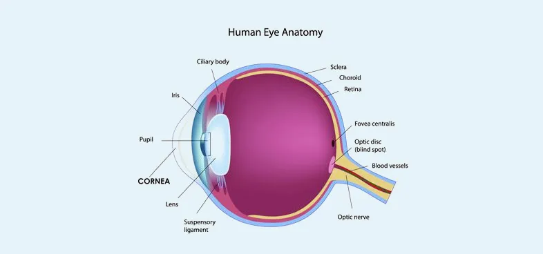
Cornea Unit
Cornea makes this world beautiful to a person. If it loses its texture and its transparency it becomes white, the person becomes blind and also makes him ugly to this world.
The cornea and ocular surface department at Lotus Eye Hospital & Institute is dedicated to the management of cornea and ocular surface disorders. The department consists of well-trained corneal surgeons and is fully equipped with the latest technology to provide state-of-the-art treatment and care for patients suffering from a wide variety of corneal disorders and infections. The department has an elaborate academic and surgical training program for post-graduate students and general ophthalmology fellows.
Diseases affecting the cornea and ocular surface:
| Microbial infections | Ocular surface disorder |
| Allergic eye diseases | Ocular surface tumors |
| Corneal dystrophies | Stevens Johnson’s syndrome |
| Keratoconus and other corneal degenerations | Chemical Injuries |
| Congenital and hereditary corneal disorders | Trauma |
Corneal dystrophies are inherited conditions where patients develop bilateral and symmetrical corneal opacities commonly involving the central cornea. This condition can cause a decrease in vision, as well as symptoms of irritation and watering.
Keratoconus is a degenerative disorder of the eye in which structural changes within the cornea cause it to thin and change to a more conical shape than its normal gradual curve.
A ‘dry eye’ is a condition where patients suffer from irritation and discomfort in the eye because of the decreased quantity of tears or increased evaporation of tears from the eye.
Microbial Infections:Bacterial, viral, fungal, and parasitic infections of the cornea may be sight-threatening and at times, serious enough to cause loss of the eye. Timely diagnosis of the pathogen and appropriate treatment can help to save the eye and restore vision. Lotus Eye Hospital and Institute's cornea department is supported by a microbiology laboratory for the isolation of the organism.
Chemical Injuries:Industrial and domestic use of chemicals is widespread. Many patients with acid and alkali injuries to the ocular surface are successfully treated at Lotus Eye Hospital and Institute hospital. Early and appropriate medical treatment followed by surgical interventions in the form of amniotic membrane grafts and autologous stem cell transplants are undertaken at Lotus Eye Hospital and Institute hospital.
Investigations
Specular Microscopy: A Specular Microscope helps to document the health of the corneal endothelium and diagnose early cases of Fuchs endothelial dystrophy. The appropriate management of cataracts with compromised corneas is also helped by specular microscopy. Keeping a track of the endothelial cell count pre and post-penetrating keratoplasty is made possible with clinical specular microscopy. Specular microscopy is also important before collagen cross-linking and phakic intraocular lens implantation.
Photo slit-Lamp: Documentation of corneal pathology is aided by the Topcon slit lamp with a digital camera unit and computer attachment. Tonopen: Intraocular pressure measurement in irregular, scarred, edematous corneas may be more accurately measured with this instrument. While applanation tonometry is still the gold standard for intraocular pressure measurement, to open provides a quick and reliable measurement in scarred, irregular corneas and post penetrating keratoplasty where there may be variable corneal thickness and astigmatism.
Corneal Topography: Lotus eye hospital & Institute has Orbscan II which can evaluate the topography of the cornea as well as to measure the corneal thickness. The orb scan has Placido and slits scanning technology which can assist in detecting forme fruste keratoconus, post Lasik ectasia, and posterior keratoconus. Planned selective suture removal in penetrating and anterior lamellar keratoplasty is made easy with this technology. Aberrometry measures the optical aberrations of the eyeball.
Ultrasonic Biomicroscopy (UBM): UBM is an important tool in scarred and hazy corneas. Angle structures, crystalline lenses, and anterior or posterior chamber intraocular lenses can be visualized in cases where there is no visibility of the anterior segment.
Anterior segment OCT: It is used to quantify the thickness of the cornea and to precisely locate the depth of corneal opacity and see the integrity of Descemet's membrane helps in lamellar keratoplasty.
Facilities
The department offers the most recent advances in the field of corneal surgery which include:
OCULAR SURFACE DISORDERS TREATMENT:
Amniotic membrane grafts and limbal cell transplant Corneal tattooing for unsightly scars in non-seeing eyes Pterygium surgery with conjunctival autografts with fibrin glue Corneal collagen crosslinking Sever Dry eye management with punctual occlusion- temporary and permanent Management of corneal erosions, recurrent epithelial defects with an anterior stromal puncture and PTK EDTA chelation for band-shaped keratopathy Bandage contact lens fitting, Research & Teaching activities, Postgraduate teaching: Aims at providing an in-depth knowledge of various corneal disorders and diagnostic procedures to the postgraduate students.
Research
Major ongoing research projects are
Start your journey to see the world better with the experts in eye care


