Overview
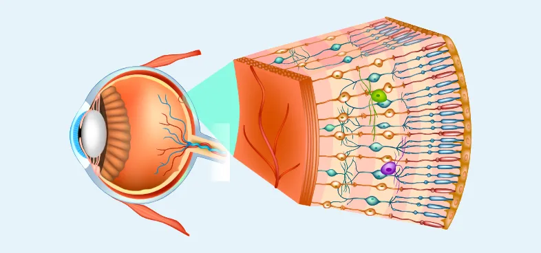
Retina is the light sensitive lining in the back of eye. It contains millions of special nerve cells which convert light to electrical impulses. These impulses are carried by the optic nerve to the brain where it is converted to images that you see.
We specialize in diagnosis and treatment of all Retinal Diseases which includes the following:
Diabetic Retinopathy
India has an enormous burden of type 2 diabetic population of nearly 50 million and it continues to rise. Abnormal blood sugar damages the blood vessels of the retina which can cause impaired vision and blindness. Diabetic vision loss occurs due to
Stages and Types of diabetic retinopathy
Symptoms
Early stages may be without any symptoms. Hence diabetic patients must regularly check their eyes with Retinal Examinations.
Common Symptoms
Tests for Diagnosing Diabetic Retinopathy
Treatment and Management of Diabetic Retinopathy
This is a specialized surgery to remove the blood within the eye and settle the retina that is being pulled away from the back of the eye. This is performed in a complicated situation.
Patient participation in their eye care is important. Patient education and also involving the primary care physician for diabetes control is the method adopted at our Institute to get the best results.
Retinal Vein Occlusion
Occlusion occurs when the vein drawing blood from the retina becomes blocked. This leads to bleeding & swelling within Retina in vein occlusion. High blood pressure, uncontrolled diabetes, and blood disorders can cause this condition. Diagnostic tests like FFA and OCT are done and intravitreal injection combined with laser help in early resolution.
Retinal Detachment / Retinal Tear
Separation of retina from its underlying layers that supply nourishment is Retinal Detachment. When a hole or tear allows fluid to pass behind the retina, it pulls off from the back surface of eye.
Some people at greater risk of detachment than the rest are patients with myopia, Lattice degeneration, Ocular Trauma, or eye surgery.
Surgery is the only treatment that can reattach the retina. Special surgeries done are,
Age Related Macular Degeneration (ARMD)
ARMD is a common cause of vision loss in elderly. This disease is often Bilateral.
Symptoms:
Types of ARMD:
There are 2 types
Tests helpful in Diagnosis are:
Treatment and progress:
Patients with ARMD require regular monitoring of their central vision with Amsters chart.
Macular edema
This occurs when fluid accumulates in macula that can impair vision.
Different causes include:
Diagnosis & Treatment:
FFA, OCT & ICG are helpful in diagnosis.
Vitreous hemorrhage
Occurs when blood collects within the vitreous. This is a jelly that fills the eye between the lens and the retina. Bleeding originates from the blood vessel of the retina which ruptures due to high blood pressure, uncontrolled diabetes, or bleeding disorders. There is a sudden severe decrease in vision. Diagnostic tests like Ultrasound B.Scan give detailed information. Treatment in the form of vitrectomy helps to remove the blood, release the traction and restore vision.
Central serous retinopathy
Occurs due to collection of fluid below center of retina called Macula.
Symptoms include:
Diagnostic Tests performed:
Treatment for CSR:
Inherited retinal diseases
Night-Blindness and many inherited retinal diseases manifest first in childhood. The retinal screening will help in identifying these diseases. Low vision aids and correction of refractive errors help the patients in managing their day-to-day activities. Special investigation helps in diagnosing disease and monitoring the rate of progression.
Myopic retinal degeneration
Retina is divided in central (called macula) & peripheral retina
Myopia causes progressive thinning of the Retina. Areas of the thinned retina can develop holes in the Retinal periphery and increase the risk of developing retinal detachment. Progressive thinning involving the central (macula) of the retina can cause a significant decrease in vision or the development of a bleeding membrane.
Patients with myopia require yearly retinal evaluation for early diagnosis and treatment of complications.
Ocular trauma
Trauma to the face and eye can be very serious and visually debilitating. People sustain ocular trauma at work, road, in traffic accidents, in sports-related, assaults, in fireworks, and due to chemical injuries. Emergency care is provided by experts. Complicated Vitreo-Retinal surgeries are performed with intraocular foreign body removal to restore vision.
Flashes & floaters
You may see flies or specks in your field of vision. This happens when vitreous degenerates or detaches. Flashes occur when Retina is pulled and it is irritated or disturbed.
Causes of Flashes & Floaters:
Detailed evaluation of the retina by a retinal specialist helps to evaluate the Retina and treat the underlying cause.
Ocular oncology
Tumors arise within the eye. They may be benign or malignant. Choroidal melanoma is the most common primary eye cancer in adults and Retinoblastoma is the most common primary intraocular tumor in children. Eyelid tumors are Basal cell carcinoma and Squamous cell carcinoma.
Detailed evaluation includes examination under anesthesia for children combined with Ultrasound B Scan. Ocular treatment like laser therapy and cryotherapy is done in collaboration with an oncologist to preserve vision and save a life.
Start your journey to see the world better with the experts in eye care


